Nobel Prize in Medicine 2026: How 3 Scientists Used Light To Switch Brain Cells On And Off
The 2026 Nobel Prize in Physiology or Medicine recognised Karl Deisseroth, Peter Hegemann and Georg Nagel for discoveries that laid the foundation of optogenetics. The technique uses light-sensitive proteins and genetic engineering to control specific nerve cells, opening new possibilities for studying brain activity, memories and potential treatments such as vision restoration.

A long-standing researchers’ dream of using a switch to control nerve cells and understand how the brain forms memories, feelings and behaviours is no longer just a dream. Awarded the 2026 Nobel Prize in Physiology or Medicine on October 5, three scientists have helped make it possible to switch the activity of individual nerve cells on or off using light.
Karl Deisseroth, 54, Peter Hegemann, 71, and Georg Nagel, 73, were recognised for discoveries that laid the foundation for optogenetics, a biological technique that uses light and genetic engineering to control the activity of specific cells.
One notable advanced application of optogenetics is the potential restoration of vision by activating nerve cells in the eyes. Visual optogenetics uses gene therapy to deliver a light-sensitive protein to cells in the retina. Light can then stimulate these cells to produce a visual signal that is sent to the brain, potentially helping restore vision in people with certain forms of visual impairment.
How 3 Researchers Turned On And Off A Nerve Cell
The foundation of this new era in neuroscience began with an alga, a plant-like organism that lives in or near water and makes its own food using sunlight. Hegemann became curious about how Chlamydomonas, a single-celled alga, reacts so rapidly to light.
Chlamydomonas has an eyespot, a tiny orange structure on its surface that contains the light-capturing molecule retinal. When Hegemann illuminated the eyespot, the alga reacted rapidly, showing an electrical impulse just half a millisecond after the light reached it.
By comparison, the human eye takes about 10 milliseconds to respond to light, involving a much more complex process.
This rapid response increased Hegemann’s curiosity. In the 1990s, he hypothesised that a single protein complex could capture light and act as an ion channel. The proposal was initially met with skepticism because, although researchers knew of several ion channels, none were known to respond directly to light.
He then attempted to isolate Chlamydomonas’s light-sensitive protein but faced difficulties because the proteins were unstable and easily damaged.
Several years later, Japanese researchers mapped the DNA of Chlamydomonas, which swims towards a light source, making the genetic codes for thousands of its genes available.
Among these genetic sequences, Hegemann’s group identified two genes that showed similarities to light-capturing proteins.
Hegemann then contacted Georg Nagel at the Max Planck Institute for Biophysics in Frankfurt to investigate whether the two genes were the ones he was looking for.
Hegemann’s Hypothesis Proved True
After collaborating with Nagel, Hegemann’s once-sceptical hypothesis gained experimental support. Nagel injected the two Chlamydomonas genes into frog eggs to investigate how the proteins worked. He found that the proteins could form ion channels that responded to light, supporting Hegemann’s hypothesis.
The Two Proteins In Human Kidney Cells
The two proteins were named channelrhodopsin-1 and channelrhodopsin-2. Like Hegemann, Nagel showed that channelrhodopsin-2 responded strongly to light. Once light was detected, a channel opened on the frog egg cell’s surface within about 0.2 milliseconds.
The researchers then tested the channelrhodopsin-2 gene in embryonic human kidney cells and hamster kidney cells. The cells produced an electrical signal when exposed to light. Their findings were published in 2003.
A Young Scientist Joins The Team
In the 1990s, Karl Deisseroth was studying medicine and considering becoming a neurosurgeon. During part of his training at a psychiatric clinic, however, he became interested in understanding patients’ suffering and their need for help.
He later earned a PhD in neuroscience and started his own research group, searching for a protein that could trigger electrical impulses in nerve cells. After extensive testing, Deisseroth wrote to Georg Nagel and asked for access to the DNA encoding the protein.
When the DNA reached Deisseroth’s lab, he introduced it into rat nerve cells grown in petri dishes. Despite concerns that the foreign gene could harm the sensitive cells, they began producing channelrhodopsin-2 without apparent problems. When exposed to blue light, the cells responded immediately, triggering nerve signals that could spread to other nerve cells.
In 2005, Deisseroth’s research group published the findings, marking a major milestone in optogenetics. However, Deisseroth knew the next challenge was to take the technique beyond lab-grown cells and use light to control nerve-cell activity in live animals.
Deisseroth then turned the discovery into a practical tool. He introduced the channelrhodopsin gene into rat nerve cells and found that blue light could switch the cells on, effectively turning channelrhodopsin into a light-controlled switch for nerve cells.
Deisseroth expanded his work through collaborations with researchers including Peter Hegemann and Georg Nagel. Researchers soon identified more light-sensitive proteins that could switch nerve cells on and off at different wavelengths. In 2006, the technique was named optogenetics.
Nerve Cells Activated In Mice
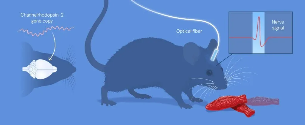
A year later, Deisseroth’s team used optogenetics to activate specific nerve cells in the brains of living mice. By introducing the channelrhodopsin-2 gene into motor-cortex neurons and stimulating them through a thin optic fibre, researchers were able to control the movement of the mice’s whiskers.
In 2007, the team also used the technique to study wakefulness. After introducing channelrhodopsin-2 into nerve cells believed to regulate arousal, researchers activated them with light and successfully woke sleeping mice.
Another major breakthrough came in 2012, when Deisseroth and Susumu Tonegawa used optogenetics to reactivate an engram-the neural pattern associated with a memory. By stimulating nerve cells linked to a fear experience, they triggered fear responses in mice, providing evidence that specific nerve cells could be linked to a particular memory.
Optogenetics Moves Towards Vision Restoration
On October 1, Dallas-based Nanoscope Therapeutics received approval from the Pharmaceuticals and Medical Devices Agency (PMDA) in Japan for an urgent review of its New Drug Application (NDA). The application seeks market approval from Japan’s Ministry of Health, Labour and Welfare (MHLW) for MOGENRY (sonpiretigene isteparvovec, MCO-010), an optogenetic gene therapy being developed to restore vision in people with inherited retinal dystrophies (IRDs).

Sahil Behl is an education journalist at Jagran with over a year of experience in journalism. Prior to joining Jagran, he worked as a Sub-Editor in NDTV’s Education department, where he was responsible for writing and editing education-related content as well as managing the department’s social media presence. At Jagran, he covers a wide range of education topics, including board examinations, school updates, admissions, and job notifications, while leveraging his editorial expertise and strong understanding of digital content strategy. Sahil holds a Bachelor’s degree in Business Administration and has also completed an eight-month certification program in Data Science. Passionate about emerging technologies, particularly artificial intelligence, he closely tracks their growing role in journalism and explores how they are transforming shaping the future of the media industry.

-1791268259963_t.webp)

