
Human Eye Structure And Functioning: The human eye is usually considered as the most valuable organ of the body and rightly so as it enables us to visualize objects. Using a naturally occurring convex lens composed of transparent living material, the human eye focuses light to allow us to view objects in our environment.
Structure of Human Eye
The human eye consists of the parts as follows:
Pupil- It is a small opening in the iris and regulates the amount of light that enters the eye. Put simply, the pupil contracts if the amount of light entering the eye is more and it expands if the amount of light is less.
Cornea- It is through the cornea that light enters the eye. As per NCERT, the cornea accounts for two-thirds of the total optical power of the eye.
Iris- The iris is the coloured membrane behind the cornea. You might be aware that the colour of the iris indicates the colour of the eye.
Also Check: The Human Eye: Anatomy, Structure, Working, Function and Defects
Retina- NCERT has defined the retina as, ‘’A transparent layer forming the inner coat of the eye, it supports the choroid layer. The rays of light, on entering the eyeball, converge and form an image on the fovea—the posterior part of the eye on retina.’’
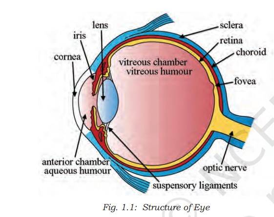
Sclera- Known as the white of the eye, the sclera is the outer covering of the eyeball. Sclera maintains the structure of the eyeball.
Lens- Lens is the transparent structure situated behind the pupil. It focuses light on the retina.
Vitreous humour- It is the gel-like substance maintaining the eyeball’s shape.
Apart from this, horizontal eye movements are controlled by the medial and lateral rectus muscles, while superior rectus and interior rectus muscles perform superior and inferior movement of the eyes.
Functioning of the Human Eye
The eye is often compared to a camera. NCERT has explained the process of how the eye captures images. ‘’The human eye is like a camera. Its lens system forms an image on a light-sensitive screen called the retina. Light enters the eye through a thin membrane called the cornea. It forms a transparent bulge on the front surface of the eyeball. The eyeball is approximately spherical in shape with a diameter of about 2.3 cm. Most of the refraction for the light rays entering the eye occurs at the outer surface of the cornea. The crystalline lens merely provides the finer adjustment of focal length required to focus objects at different distances on the retina. We find a structure called iris behind the cornea. Iris is a dark muscular diaphragm that controls the size of the pupil. The pupil regulates and controls the amount of light entering the eye. The eye lens forms an inverted real image of the object on the retina. The retina is a delicate membrane having an enormous number of light-sensitive cells. The light-sensitive cells get activated upon illumination and generate electrical signals. These signals are sent to the brain via the optic nerves. The brain interprets these signals, and finally, processes the information so that we perceive objects as they are.’’


-Answer-Keys-2026_-Download-Class-wise-OMR-Sheet-PDF-1766142098617.jpg)

Comments
All Comments (0)
Join the conversation